研究のご紹介

足の外科リサーチ

原因・病態が未解明の足の病気の研究と、治療法の取り組みを紹介します。

研究概要

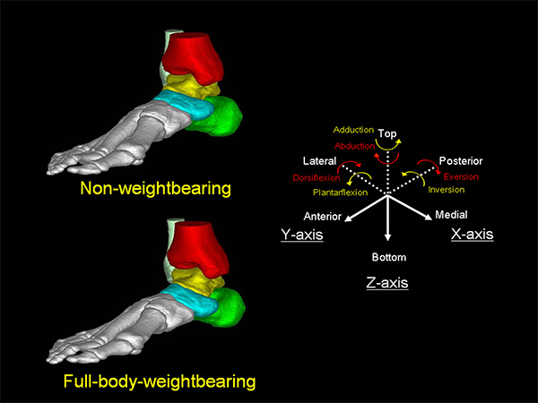
ヒトの祖先が二足直立歩行を獲得したのは、およそ1,000万年前と言われています。ヒトの足は特殊な進化をとげ、最大の特徴として縦方向と横方向にアーチ構造をもち、二足直立で歩行する際にも荷重が支持できるようになったといわれています。

足部に何らかの障害が生じた場合、正確な診断を行うには診察や画像読影に多くの知識と技術が必要です。足の外科では多様な足部障害の患者さんが受診するため、その知識と技術を身につける事が可能なだけでなく、荷重支持機構の病態を解明し,効果的な機能再建法の確立を目指して研究を行っています。

CT画像を用いた足・足関節のバイオメカニクス

CT画像から生体の三次元足根骨モデルを構築し、足関節の運動および荷重にともなう各足根骨の動きをコンピューター上で計測しました。これまで、扁平足や変形性関節症において足根骨の動きに異常があることを解明し、足底挿板などの治療の有効性を証明してきました。バイオメカニクスを応用した生体足における荷重支持機構の研究を継続しています。


後足部撮影法の開発と後足部アライメントの評価

後足部アライメント評価法として、新たな撮影法と脛骨に対する踵骨のアライメント(後足部アライメント)の計測法を開発し、実用化しました。この撮影法を用いて、人工膝関節全置換術(TKA)の後足部アライメントに対する影響を調査し、TKA術前後に後足部アライメントが経時的に変化することを明らかにして報告しています。

成人期扁平足の病態解明と治療法の開発

成人期扁平足は近年増加傾向にあり、今後も増加が予想されます。とくに後脛骨筋腱機能不全症(PTTD)は成人期扁平足の主な原因で、後脛骨筋腱の損傷により扁平足変形が進行するとされています。われわれはMRIから、後脛骨筋腱と足部靱帯の損傷と扁平足変形の進行との関連を調査しています。また、CT画像から扁平足の有限要素法を用いたモデルを作製し、各症例に応じた手術法(テーラーメイド手術法)の開発を行っています。

今後の研究

  • 凹足変形の病態解明
  • 変形性足関節症における足根骨の形状および動態の変化
  • アキレス腱のバイオメカニクス
  • 体外衝撃波治療とその応用

主な業績

  • The influence of hallux valgus and flatfoot deformity on metatarsus primus elevatus: A radiographic study.
    Kido M, Ikoma K, Sotozono Y, Ikeda R, Imai K, Maki M, Ohashi S, Kubo T.
    J Orthop Sci 25: 291-296, 2020
  • Early stage and small lesions of medial osteochondral lesions of the talus in the presence of chronic lateral ankle instability: a retrospective study.
    Ikoma K, Kido M, Maki M, Imai K, Hara Y, Ikeda R, Ohashi S, Shirai T, Kubo T.
    J Orthop Sci 25: 178-182, 2020
  • Reproducibility of radiographic methods for assessing longitudinal tarsal axes Part 1: Consecutive case study.
    Kido M, Ikoma K, Ikeda R, Hosokawa T, Hara Y, Imai K, Maki M, Ohashi S, Mikami Y, Kubo T.
    Foot (Edinb) 40: 1-7, 2019
  • Reproducibility of radiographic methods for assessing longitudinal tarsal axes Part 2: Severe cavus or flatfoot study.
    Kido M, Ikoma K, Ikeda R, Hosokawa T, Hara Y, Imai K, Maki M, Ohashi S, MikamiY, Kubo T.
    Foot (Edinb). 42:101631. 2019
  • Two-stage correction using the Taylor Spatial Frame for severe hindfoot deformity in a patient with Marfan syndrome: A case report.
    Ohashi S, Ikoma K, Kido M, Oka Y, Kim WC, Kubo T.
    J Orthop Sci 24: 178-183, 2019
  • A kinematic and kinetic analysis of the hip and knee joints in patients with posterior tibialis tendon dysfunction; comparison with healthy age-matched controls.
    Maeda H, Ikoma K, Toyama S, Taniguchi D, Kido M, Ohashi S, Kubo S, Hishikawa N, Sawada K, Mikami Y, Kubo T.
    Gait Posture 66: 228-235, 2018
  • Subcutaneous rupture of the flexor hallucis longus tendon at the musculotendinous junction in a soccer player.
    Hosokawa T, Ikoma K, Kido M, Hara Y, Maki M, Ohashi S, Kubo T.
    J Orthop Sci 14: 1-4, 2018
  • Towards patient-specific medializing calcaneal osteotomy for adult flatfoot: a finite element study.
    Wang Z, Kido M, Imai K, Ikoma K, Hirai S.
    Comput Methods Biomech Biomed Engin 21: 332-343, 2018
  • Extracorporeal shock wave therapy for adolescent patient with calcific tendinopathy of posterior tibial tendon: A case report.
    Ikoma K, Hara Y, Maki M, Ohashi S, Ueshima K, Kubo T.
    Prog Rehabil Med. 2018; 3: 20180004
  • Magnetic resonance imaging findings of chronic plantar fasciitis before and after extracorporeal shock wave therapy.
    Maki M, Ikoma K, Kido M, Hara Y, Sawada K, Ohashi S, Kubo T.
    Foot (Edinb) 33: 25-28, 2017 
  • Relationship Between Grading With Magnetic Resonance Imaging and Radiographic Parameters in Posterior Tibial Tendon Dysfunction.
    Ikoma K, Hara Y, Kido M, Imai K, Maki M, Ohashi S, Kubo T.
    J Foot Ankle Surg 56: 718-723, 2017
  • Examination of the correlation between foot morphology measurements using pedography and radiographic measurements.
    Inui K, Ikoma K, Imai K, Ohashi S, Maki M, Kido M, Hara Y, Oka Y, Fujiwara H, Kubo T.
    J Foot Ankle Surg 56: 298-303, 2017
  • Recurrence of deformity after silicone implant and resection arthroplasty of the metatarsophalangeal joint for rheumatoid arthritis: long-term results.
    Taniguchi D, Oda R, Ikoma K, Toyama S, Takatori R, Imai K, Hara Y, Sawada K, Tokunaga D, Fujiwara H, Kubo T.
    Mod Rheumatol 27: 266-270, 2017
  • Diagnostic Characteristics of Standard Radiographs and Magnetic Resonance Imaging of Ruptures of the Tibialis Posterior Tendon.
    Ikoma K, Ohashi S, Maki M, Kido M, Hara Y, Kubo T.
    J Foot Ankle Surg 55: 542-546, 2016
  • Weight-bearing three-dimensional computed tomography analysis of the forefoot in patients with flatfoot deformity.
    Yoshioka N, Ikoma K, Kido M, Imai K, Maki M, Arai Y, Fujiwara H, Tokunaga D, Inoue N, Kubo T.
    J Orthop Sci 21: 154-158, 2016
  • Non-osseous Tarsal Coalition of the Lateral Cuneocuboid Joint: A Case Report.
    Imai K, Ikoma K, Kido M, Maki M, Arai Y, Fujiwara H, Kubo T.
    J Foot Ankle Surg 55:1072-1075, 2016
  • Hindfoot alignment at one year after total knee arthroplasty.
    Takenaka T, Ikoma K, Ohashi S, Arai Y, Hara Y, Ueshima K, Sawada K, Shirai T, Fujiwara H, Kubo T.
    Knee Surg Sports Traumatol Arthrosc 24: 2442-2446, 2016
  • Joint space width of the tibiotalar joint in the healthy foot.
    Imai K, Ikoma K, Kido M, Maki M, Fujiwara H, Arai Y, Oda R, Tokunaga D, Inoue N, Kubo T.
    J Foot Ankle Res 8: 26, 2015
  • Biomechanical Effect of Augmented Soft Tissue Mobilization Therapy on Achilles Tendinitis in the Rabbit Model.
    Imai K, Ikoma K, Chen Q, Zhao CF, An KN, Gay RE.
    J Manipulative Physiol Ther 38: 112-118, 2015
  • Correlation between the outcome of extracorporeal shock wave therapy and pretreatment MRI findings for chronic plantar fasciitis.
    Maki M, Ikoma K, Imai K, Kido M, Hara Y, Arai Y, Fujiwara H, Kubo T.
    Mod Rheumatol 25: 427-430, 2015
  • Alternation of hindfoot alignment after total knee arthroplasty using a novel hindfoot alignment view.
    Hara Y, Ikoma K, Arai Y, Ohashi S, Maki M, Kubo T.
    J Arthroplasty 30: 126-129, 2015
  • Hindfoot endoscopic exision of ankle posterior impingement including elderly patients.
    Ikoma K, Maki M, Imai K, Kido M, Taniguchi D, Oda R, Fujiwara H, Kubo T.
    Clin Res Foot Ankle 2: 145, 2014
  • Effect of therapeutic insoles on the medial longitudinal arch in patients with flatfoot deformity: a three-dimensional loading computed tomography study.
    Kido M, Ikoma K, Hara Y, Imai K, Maki M, Ikeda T, Fujiwara H, Tokunaga D, Inoue N, Kubo T.
    Clin Biomech (Bristol, Avon) 29: 1095-1098, 2014
  • Extra-articular dorsal closing-wedge osteotomy to treat late-stage Freiberg disease using polyblend sutures: technical tips and clinical results.
    Ikoma K, Maki M, Kido M, Imai K, Arai Y, Fujiwara H, Mikami Y, Kubo T.
    Int Orthop 38: 1401-1405, 2014
  • A finite element model of flatfoot (Pes Planus) for improving surgical plan.
    Wang Z, Imai K, Kido M, Ikoma K, Hirai S.
    Conf Proc IEEE Eng Med Biol Soc 2014: 844-847, 2014
  • Achilles tendon lengthening for equinus foot with Miyoshi myopathy: a case report.
    Ikoma K, Maki M, Kido M, Arai Y, Fujiwara H, Kubo T.
    J Foot Ankle Surg 53: 643-646, 2014
  • A new radiographic view of the hindfoot.
    Ikoma K, Noguchi M, Nagasawa K, Maki M, Kido M, Hara Y, Kubo T.
    J Foot Ankle Res 6: 48, 2013
  • Load response of the medial longitudinal arch in patients with flatfoot deformity: in vivo 3D study.
    Kido M, Ikoma K, Imai K, Tokunaga D, Inoue N, Kubo T.
    Clin Biomech (Bristol, Avon) 28: 568-573, 2013
  • Tarsal tunnel syndrome in hemodialysis patients: a case series.
    Imai K, Ikoma K, Imai R, Fujiwara H, Kido M, Hara Y, Ono T, Kubo T.
    Foot Ankle Int 34: 439-444, 2013
  • Comparison of the Relative Bone Position Between Normal and Flat Feet Using 3-Dimensional Computed Tomography Reconstruction Images of Tarsal Bones: a Trans-sectional Research.
    Imai K, Ikoma K, Maki K, Kido M, Takatori R, Tokunaga D, Kubo T.
    Jpn J Arth 32: 463-469, 2013
  • Features of hindfoot 3D kinetics in flat foot in ankle-joint maximal dorsiflexion and plantarflexion.
    Imai K, Ikoma K, Maki M, Kido M, Tsuji Y, Takatori R, Tokunaga D, Inoue N, Kubo T.
    J Orthop Sci 16: 638-643, 2011
  • Load response of the tarsal bones in patients with flatfoot deformity: in vivo 3D study.
    Kido M, Ikoma K, Imai K, Maki M, Takatori R, Tokunaga D, Inoue N, Kubo T.
    Foot Ankle Int 32: 1017-1022, 2011