脊椎研究班は、3つのテーマを重点的に研究しています。
脊椎・脊髄リサーチ
脊椎と脊髄の疾患の新しい治療法の取り組みを紹介します。
研究概要
低侵襲手術の開発
低侵襲除圧術
われわれが考案した低侵襲除圧術である筋肉温存型椎弓間除圧術(MILD)、内視鏡下正中進入除圧術(ME-MILD)は全国的にも認知され、さまざまな施設で施行され良好な治療成績が報告されています。
Hatta Y, et al. Spine, 2009.
Mikami Y, et al. Eur Spine J, 2013.
Nagae M, et al. J Spine Res, 2015.
Tonomura H, et al. J Spinal Disdrd Tech, in press
Mori G, et al. J Neurosurg Spine, in press
側方経路腰椎椎体間固定術における手術支援機器
近年注目されている最小侵襲脊椎安定術(MISt)の1つである側方経路腰椎椎体間固定術:XLIF、OLIFを安全に行うための手術支援機器の開発に取り組んでいます。
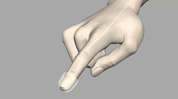
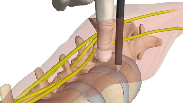
神経合併症を予防するための指電極システムの開発
Narita W, et al. J Neurosurg Spine, in press
椎間板変性に対する治療法の開発
自家多血小板血漿(PRP)の投与が家兎椎間板変性モデルで椎間板の再生に有効であることを証明し、継続して椎間板変性のメカニズムについて研究しています。椎間板変性を抑制する因子として肝細胞増殖因子(HGF)に注目し、培養椎間板細胞でその有用性を証明しました。現在は生体内での治療効果を検討しています。
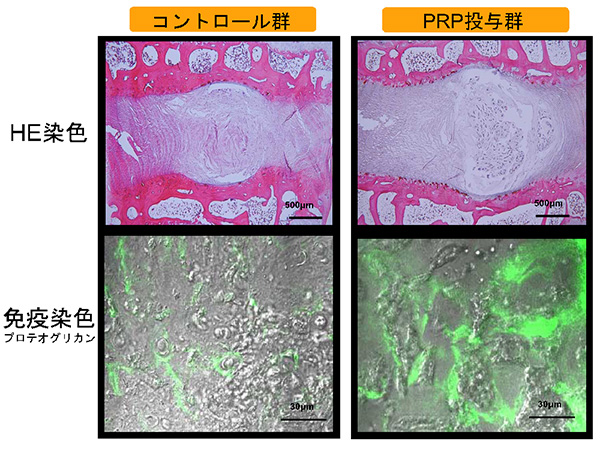
家兎椎間板変性モデルにPRPを投与し、変性抑制効果があることを確認した
Nagae M, et al. Tissue Eng. 2007.
Sawamura K, et al. Tissue Eng Part A. 2009.
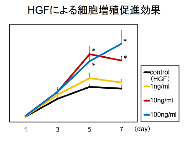
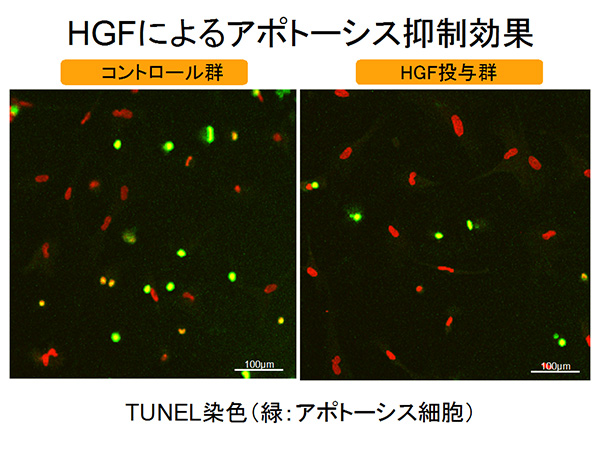
HGFは椎間板細胞の細胞増殖を促進しアポトーシスを抑制することを確認した
Ishibashi H, et al. J Orthop Res, in press.
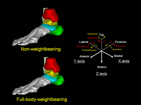
生体内三次元画像を用いた病態解析
臨床との架け橋となる生体力学的研究を教室の同門である米国Rush医科大学 井上 望先生の研究室と協力して行っています。
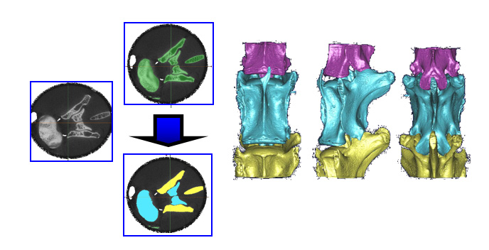
三次元画像を用いたリウマチ頚椎の病態解析
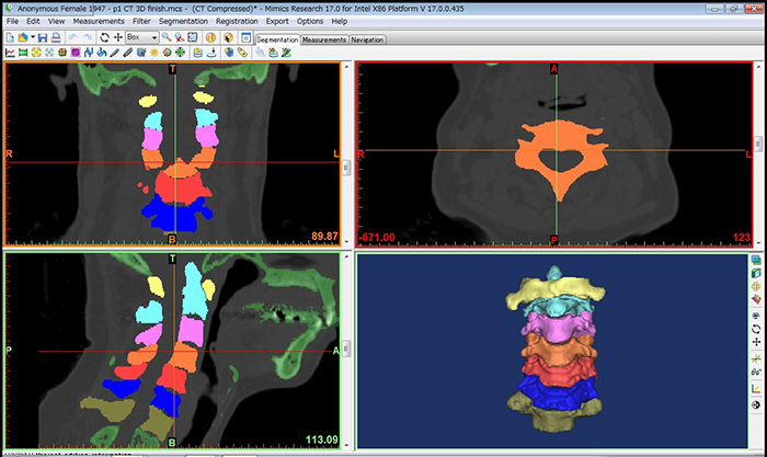

リウマチ頚椎三次元画像モデル
Takatori R, et al. Clin Exp Rheumatol, 2008.
Takatori R, et al. Spine, 2010.
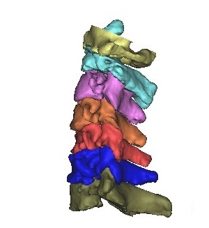
Micro CTを用いた動物実験での三次元画像解析
家兎三次元画像モデル(Micro CT)
主な業績
- Hepatocyte growth factor promotes nucleus pulposus cell proliferation with HIF-1α expression
Itsuji T, Tonomura H, Ishibashi H, Oyabu K, Takatori R, Nagae M, Mikami Y, Tanida T, Tanaka M, Kubo T
J Orthop Res (Online ahead of print) - Can relatively large cervical spinal cord for spinal canal reflect severity of paralysis in elderly patients of cervical spinal cord injury caused by minor trauma?
Koike H, Hatta Y, Tonomura H, Nonomura M, Takatori R, Mikami Y, Nagae M, Kubo T
Medicine (Baltimore), 26: e20929, 2020 - The Potential Role of Hepatocyte Growth Factor in Degenerative Disorders of the Synovial Joint and Spine
Tonomura H, Nagae M, Takatori R, Ishibashi H, Itsuji T, Takahashi K
Int J Mol Sci, 18: 8717, 2020 - Leg muscle strength after lateral interbody fusion surgery recovers over time after temporary muscle weakness
Takatori R, Ogura T, Narita W, Hayashida T, Tanaka K, Tonomura H, Nagae M, Mikami Y, Kubo T
Clin Spine Surg, 32: E160-E165, 2019. - Clinical outcome of muscle-preserving interlaminar decompression(MILD)for lumbar spinal canal stenosis: minimum 5-year follow-up study
Hatta Y, Tonomura H, Nagae M, Takatori R, Ikeda T, Mikami Y, Kubo T
Spine Surg Relat Res, 29: 54-60, 2018. - Facet joint osteoarthritis affects spinal segmental motion in degenerative spondylolisthesis
Kitanaka S, Takatori R, Arai Y, Nagae M, Tonomura H, Mikami Y, Inoue N, Ogura T, Fujiwara H, Kubo T
Clin Spine Surg, 31: E386-E390, 2018. - Posterior resection of fifth lumbar giant schwannoma combined with a recapping transiliac approach: case report and technical note
Tonomura H, Hatta Y, Nagae M, Takatori R, Kubo T
Eur J Orthop Surg Traumatol, 28: 1209-1214, 2018. - Osteoporotic effect on bone repair in lumbar vertebral body defects in a rat model
Sakata M, Tonomura H, Itsuji T, Ishibashi H, Takatori R, Mikami Y, Nagae M, Matsuda KI, Tanaka M, Kubo T
J Orthop Surg(Hong Kong), 26: 2309499018770340, 2018. - Platelet-rich plasma combined with gelatin beta-tricalcium phosphate sponge promote bone regeneration of vertebral body defects in a rat osteoporosis model
Sakata M, Tonomura H, Itsuji T, Ishibashi H, Takatori R, Mikami Y, Nagae M, Matsuda K, Tabata M, Tanaka M, Kubo T
Tissue Eng Part A, 24: 1001-1010, 2018. - Effect of three-dimensional rotational deformity correction in surgery for adult degenerative scoliosis using lumbar lateral interbody fusion and posterior pedicle screw fixation
Takatori R, Ogura T, Narita W, Hayashida T, Tanaka K, Tonomura H, Nagae M, Mikami Y, Kubo T
Spine Surgery and Related Research, 2(1): 65-71, 2018 - A new method of measuring the occipitocervical angle that could be applied as an intraoperative indicator during occipitocervical fusion
Nagashima S, Nagae M, Arai Y, Tonomura H, Takatori R, Sukenari T, Fujiwara H, Mikami Y, Kubo T
Clin Spine Surg, 30(7):E981-E987, 2017 - Magnetic resonance imaging evaluation of the effects of surgical invasiveness on paravertebral muscles following muscle-preserving interlaminar decompression (MILD)
Tonomura H, Hatta Y, Mikami Y, Ikeda T, Harada T, Nagae M, Koike H, Hase H, Kubo T
Clin Spine Surg, Mar, 30(2):E76-E82, 2017 - Innovative technique for the placement of the drainage tube for microendoscopic spinal decompression
Mizuno K, Mikami Y, Hase H, Ikeda T, Nagae M, Tonomura H, Shirai T, Fujiwara H, Kubo T
Clin Spine Surg, Feb;30(1):E59-E63, 2017 - Usefulness of anterior cervical fusion with titanium interbody cage for treatment of cervical degenerative with disease preoperative segmental kyphosis
Hosoi K, Tonomura H, Takatori R, Nagae M, Mikami Y, Arai Y, Fujiwara H, Kubo T
Medicine (Baltimore), Aug;96(32):e7749, 2017 - A case of bone cement used for spinal reconstruction after lumbosacral vertebral tumor excision extravasating and penetrating the gastrointestinal tract
Nagae M, Mikami Y, Mizuno K, Harada T, Ikeda T, Tonomura H, Fujiwara H, Kubo T
Medicine (Baltimore), Oct;95(42):e5178, 2016 - Prevention of neurological complications using a neural monitoring system with a finger electrode in the XLIF approach
Narita W, Takatori R, Arai Y, Nagae M, Tonomura H, Hayashida T, Ogura T, Fujiwara H, Kubo T
J Neurosurg Spine, Oct;25(4):456-463, 2016 - Outcomes in cases of lumbar degenerative spondylolisthesis more than 5 years after treatment with minimally invasive decompression: Examination of pre- and postoperative slippage, intervertebral disc changes and clinical results
Mori G, Mikami Y, Arai Y, Ikeda T, Nagae M, Tonomura H, Takatori R, Sawada K, Fujiwara H, Kubo T
J Neurosurg Spine, Mar;24(3):367-74, 2016 - Hepatocyte growth factor/c-Met promotes proliferation, suppresses apoptosis and improves matrix metabolism in rabbit nucleus pulposus cells in vitro
Ishibashi H, Tonomura H, Ikeda T, Nagae M, Sakata M, Fujiwara H, Tanida T, Matsuda KI, Kawata M, Kubo T
J Orthop Res, Apr;34(4):709-16, 2016